Introduction
In neurological practice, some of the simplest checks still carry a lot of value. Pupillary evaluation is one of them. It is usually done early in the neuro exam and repeated whenever there is a concern about changes in neurological status.
At a basic level, clinicians are looking at two things: size and reaction to light. Even this simple observation can give clues about how certain neurological pathways are functioning. That is why pupil measurement continues to be part of routine assessment, whether the setting is emergency care, ICU, or general neurology.
While the approach has stayed largely the same, the way it is documented and interpreted has slowly changed with the use of neurological tools.
Where Pupillary Evaluation Fits in the Neuro Exam
During a neuro exam, pupillary evaluation helps assess pathways that involve the optic nerve and brainstem. A normal response is fairly straightforward. The pupil constricts when light is applied and then returns to its baseline size.
What matters more is when the response is not typical. A slow reaction, unequal pupils, or no response at all can point toward a neurological issue. These findings are not used alone, but they often guide what needs to be checked next.
In patients who are not fully responsive, pupil measurement becomes even more useful. Other parts of the neuro exam may be difficult to perform, but pupil response can still be assessed with some consistency.
How Pupillary Evaluation Is Done
Most clinicians still rely on a penlight for pupillary evaluation. It is quick, requires no setup, and works in almost any setting. The clinician observes how the pupil reacts and notes the findings.
The limitation is that this method depends heavily on observation. Lighting conditions, examiner experience, and even how the test is performed can influence what is seen. In busy settings, the exam is often brief, which makes subtle differences harder to pick up.
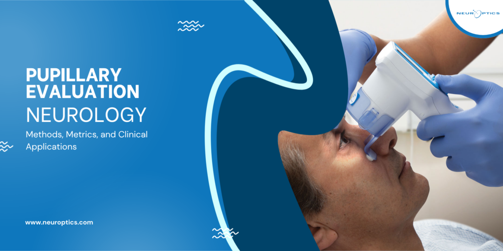
To reduce this variation, many settings now use neurological tools that provide objective pupil measurement. These devices deliver a consistent light stimulus and record how the pupil responds. The output is numerical, which makes it easier to document and compare.
This does not replace visual assessment, but it does add a level of consistency that can be helpful when exams are repeated.
Understanding What Gets Measured
When using objective pupil measurement, several values are usually recorded. These include the starting size of the pupil, how quickly it constricts, how it returns to baseline, and how much it changes overall.
Each of these reflects a slightly different part of the response. Looking at them together gives a clearer picture than relying on a single observation.
Many neurological tools also provide an NPi, which combines these responses into one value. The NPi is easier to track over time, but it is still based on the same underlying measurements. It works best when used along with those individual values rather than instead of them.
Why It Matters in Clinical Practice
In day-to-day practice, pupillary evaluation is not done in isolation. It is part of ongoing monitoring, especially in patients where neurological status can change.
In emergency settings, a quick pupil measurement may be one of the first neurological indicators available. In ICU settings, repeated checks allow clinicians to look at patterns instead of single readings.
There are also situations where the rest of the neuro exam is limited. Sedation, intubation, or reduced consciousness can make it difficult to assess other responses. In those cases, pupil response becomes one of the few things that can still be followed over time.
Using the same neurological tools repeatedly helps make these comparisons more reliable.
Consistency and Documentation
One challenge with traditional pupillary evaluation is how findings are recorded. Terms like reactive or sluggish can mean different things to different clinicians.
With structured pupil measurement, the findings are clearer. Numbers are easier to compare than descriptions, especially when multiple clinicians are involved in care.
During handovers, this becomes important. Instead of interpreting written descriptions, incoming clinicians can look at previous values and understand what has changed.
Consistency in how the exam is performed and documented makes a noticeable difference over time.
Final Thoughts
Pupillary evaluation is still one of those basic checks that clinicians rely on regularly. It doesn’t take much time, but it often adds useful context, especially when other findings are limited.
As neurological tools are used more often, pupil measurement is being recorded in a more structured way. Values like the NPi can help when looking at changes over time, but they don’t stand on their own.
When considered along with the rest of the neuro exam, these measurements become more meaningful. They support ongoing assessment, but they don’t replace clinical judgment.