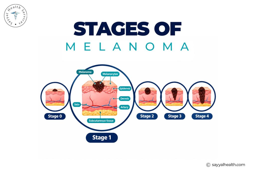
Receiving a skin cancer diagnosis can feel overwhelming, but understanding the precise stage of the condition provides clarity on prognosis and treatment pathways. Cutaneous melanoma—a malignancy originating in the pigment-producing cells (melanocytes) of the skin—is categorized across five primary numerical stages, beginning at Stage 0 and progressing through Stage IV.
Among these classifications, melanoma in situ represents the earliest detectable form of the disease. Caught at this pre-invasive boundary, the abnormal cells are entirely localized within the outer layer of the skin, offering an exceptionally high cure rate.
Understanding how clinicians stage skin tumors, what differentiates non-invasive lesions from invasive disease, and how treatment shifts across stages empowers patients to make informed healthcare decisions.
What Is Melanoma in Situ (Stage 0)?
“In situ” is a Latin phrase translating to “in its original place.” In dermatopathology, melanoma in situ (Stage 0) means that malignant melanocytes are confined exclusively to the epidermis—the top layer of skin.
Because the epidermis lacks blood vessels and lymphatic channels, cancer cells at Stage 0 cannot travel through the vascular or lymphatic systems to distant organs.
Key Characteristics of Stage 0:
- Cellular Location: Restricted strictly to the epidermal layer above the basement membrane.
- Breslow Depth: 0.0 mm (no invasion into the deeper dermis).
- Metastatic Risk: Zero to negligible risk of regional or distant spread.
- Primary Treatment: Surgical excision with clear anatomical margins.
The American Joint Committee on Cancer (AJCC) Staging Framework
Skin cancer staging follows the standardized TNM system developed by the AJCC. Staging takes into account three core variables:
- Tumor (T): Thickness of the primary lesion in millimeters (Breslow depth) and presence of ulceration (breakdown of the skin layer over the tumor).
- Nodes (N): Spread of cancer cells to nearby regional lymph nodes.
- Metastasis (M): Dissemination of tumor cells to distant skin sites, lungs, brain, or abdominal organs.
Breakdown of Melanoma Stages: From Stage 0 to Stage IV
Each stage reflects the extent to which cancer cells have grown locally or spread throughout the body.
Stage 0 (Melanoma in Situ)
Non-invasive, flat or irregular pigmented macule confined to the epidermis.
Stage I (Early Invasive Localized Disease)
The tumor has invaded the dermis but remains small and localized without spreading to lymph nodes.
- Stage IA: Tumor thickness is less than 0.8 mm without ulceration.
- Stage IB: Tumor thickness is between 0.8 mm and 1.0 mm without ulceration, or less than 0.8 mm with ulceration.
Stage II (High-Risk Localized Disease)
The primary tumor is thicker (greater than 1.0 mm to over 4.0 mm) and may exhibit ulceration, indicating a higher risk of microscopic spread, but lymph nodes remain clear.
For a detailed visual guide and breakdown of clinical classifications across localized, regional, and distant disease, review the Sayyal Health guide to melanoma stages, which outlines the diagnostic criteria doctors use during patient consultations.
Stage III (Regional Lymph Node Involvement)
Cancer cells have spread beyond the primary skin site into regional lymph channels, sentinel lymph nodes, or surrounding in-transit tissue.
Stage IV (Distant Metastatic Disease)
Malignant cells have metastasized through the bloodstream to distant skin areas, lymph nodes, or organs such as the lungs, liver, brain, or gastrointestinal tract.
Melanoma Staging Overview Table
| Stage | Classification | Breslow Depth / Clinical Extent | Regional Lymph Nodes | 5-Year Relative Survival Rate* |
| Stage 0 | Melanoma in Situ | 0.0 mm (Epidermis only) | Negative | ~99% – 100% |
| Stage I | Localized (Thin) | $\le 1.0$ mm | Negative | ~97% – 99% |
| Stage II | Localized (Thick) | $> 1.0$ mm to $> 4.0$ mm | Negative | ~82% – 93% |
| Stage III | Regional Spread | Any depth; presence of skin ulceration variable | Positive (Nodal involvement) | ~60% – 78% |
| Stage IV | Distant Metastasis | Any depth | Positive or Negative | ~25% – 35% |
How Is Melanoma Staged Clinically?
Accurate staging requires a multi-step diagnostic workup:
- Excisional Biopsy: The suspicious lesion is surgically removed and examined under a microscope by a dermatopathologist to determine the exact Breslow thickness, mitotic rate, and ulceration status.
- Sentinel Lymph Node Biopsy (SLNB): Recommended for lesions thicker than 0.8 mm (or thinner lesions with ulceration). A radioactive tracer and dye pinpoint the first draining lymph node to check for microscopic tumor cells.
- Diagnostic Imaging: PET-CT scans, brain MRIs, and chest CTs are utilized for high-risk Stage II or Stage III/IV patients to evaluate potential internal metastasis.
- Genetic / Biomarker Testing: Tumors from advanced stages undergo testing for genetic mutations—such as BRAF V600E—to select targeted molecular therapies.
Treatment Approaches by Stage
Medical management depends on the confirmed stage at the time of diagnosis.
- Stage 0 (In Situ): Treated with wide local excision (WLE), removing the lesion along with a safe margin of healthy tissue (typically 5 mm to 10 mm) under local anesthesia.
- Stages I & II: Surgical excision with wider surgical margins (1 cm to 2 cm). High-risk Stage II cases may receive adjuvant systemic therapies to decrease recurrence risks.
- Stage III: Wide excision of the primary site alongside surgical removal of affected lymph nodes, followed by targeted drug therapy (BRAF/MEK inhibitors) or immune checkpoint inhibitors (PD-1 inhibitors like pembrolizumab or nivolumab).
- Stage IV: Managed primarily via systemic immunotherapy, dual checkpoint blockade, targeted therapy, radiation, or participation in clinical trials.
Frequently Asked Questions
Can melanoma in situ come back after surgery?
Recurrence after complete surgical removal of Stage 0 melanoma with clear margins is rare (less than 5%). However, individuals diagnosed with one melanoma have a higher risk of developing new, separate primary skin cancers elsewhere on their body.
Is a sentinel lymph node biopsy needed for Stage 0 melanoma?
No. Because melanoma in situ is strictly confined to the epidermis and lacks access to lymphatic vessels, a sentinel lymph node biopsy is not clinically indicated for Stage 0 lesions.
What is the ABCDE rule for identifying suspicious skin spots?
The ABCDE mnemonic helps identify potential skin lesions that warrant professional evaluation:
- A – Asymmetry: One half of the spot does not match the other.
- B – Border: Edges are irregular, ragged, notched, or blurred.
- C – Color: Color is uneven and may include shades of brown, black, pink, red, or white.
- D – Diameter: The spot is larger than 6 mm across (about the size of a pencil eraser), though some can be smaller.
- E – Evolving: The spot changes in size, shape, color, or elevation over time.
How often should someone with a history of melanoma get skin checks?
Patients with a history of melanoma should undergo full-body professional dermatological exams every 3 to 6 months for the first 2 years, followed by bi-annual or annual skin checks for life, alongside monthly self-examinations.
Summary
Early detection remains the single most effective tool in managing skin malignancies. Diagnosing a lesion at Stage 0 (melanoma in situ) means treatment can be completed with a minor outpatient surgical procedure, yielding exceptional long-term outcomes. By performing regular skin self-exams, protecting skin from excessive ultraviolet (UV) exposure, and seeking prompt evaluation for changing moles, individuals can ensure skin changes are identified at their earliest, most treatable stages.